State-of-the-art diagnostic technology designed to detect, monitor, and protect your vision with precision. Technological advancements have transformed the way eye diseases are detected, monitored, and treated. Our office continually invests in the latest innovations to improve diagnostic accuracy, enhance patient comfort, and deliver the highest standard of care.
Eye Care Technology
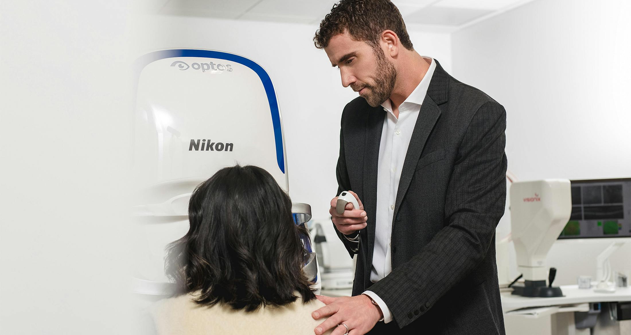

Utilizing Advanced Technology to Care for Your Eyes
Technological advancements have transformed the way eye diseases are detected, monitored, and treated. Our office continually invests in the latest innovations to improve diagnostic accuracy, enhance patient comfort, and deliver the highest standard of care.
Myopia Management Technology Myopia Master® by OCULUS
We are proud to offer the cutting-edge Myopia Master, new to Richlin & Associates—and one of the first in Los Angeles, which combines:
- Auto-refraction
- Corneal curvature measurement (keratometry)
- Axial length measurement
- Integrated myopia management software
This advanced system allows for earlier and more reliable detection of myopia (nearsightedness), leading to more accurate diagnoses and timely treatment—especially important as myopia becomes increasingly prevalent in children. The Myopia Master is an integral part of our Myopia Control Center. If your child experiences blurry distance vision, we recommend scheduling an evaluation. Visit Myopia Control Center

Schedule A Consultation
Contact UsDry Eye Treatment Technology LipiFlow® Thermal Pulsation
LipiFlow is an advanced treatment for Meibomian Gland Dysfunction (MGD), a leading cause of dry eye disease. Studies show that approximately 86% of dry eye patients have underlying MGD. This quick, drug-free treatment:
- Improves gland function
- Is clinically proven in peer-reviewed studies
- Provides long-term symptom relief

Diagnostic & Functional Testing
Peripheral Vision Testing
We utilize the Humphrey Visual Field Analyzer, the gold standard for detecting peripheral vision loss and diagnosing glaucoma and neurological conditions.
For screening, we use Frequency Doubling Technology (FDT) for rapid early detection. For advanced cases, we use the Humphrey-Zeiss HFA analyzer for comprehensive visual field analysis.
Corneal Topography
Using the Medmont E300 Corneal Topographer, we map the curvature of the cornea with extreme precision. Since the cornea contributes approximately 70% of the eye’s refractive power, accurate mapping is essential for:
- Specialty contact lens fitting
- Orthokeratology
- Keratoconus management
- Refractive surgery planning
Digital Imaging
Our Huvitz ReSeeVit HR Elite Digital Camera captures detailed images of the front surface of the eye, allowing us to document and monitor conditions such as:
- Corneal ulcers
- Viral infections
- Foreign bodies
Digital Refractors
The Huvitz HDR-7000 Digital Refractor allows us to precisely measure refractive error and determine your best-corrected vision. It also assists in identifying astigmatism and eye muscle imbalance (strabismus).

Office Tour